
The Difference the Right Tools Can Make
Generous 2018 Gift from Alumnus Thomas Dudash Enables Foundational MEMS Research
PITTSBURGH (Jan. 16, 2019) — Sometimes, in order to understand the big picture, you need to start by assessing the smallest of details. It’s a truth that engineers know well — selecting the right materials can mean the success or failure of a given application.
As technology advances, researchers have assessed engineering materials at the microscopic level for applications ranging from nanomachines to semiconductors, specialized coatings to robotics. For researchers at the University of Pittsburgh’s Swanson School of Engineering, looking closely enough to engineer materials for cutting-edge applications would not have been possible without the generous $1 million gift that Thomas F. Dudash provided in 2018.
Mr. Dudash, an alumnus of the University of Pittsburgh who received his bachelor’s degree in metallurgical engineering in 1960, never imagined that he’d have a million dollars to donate for advanced research. After a lifelong career with Allegheny Ludlum, he wanted to share his success with the next generation of materials engineers.
The gift was designated for the Department of Mechanical Engineering and Materials Science (MEMS), the successor to the metallurgical engineering program. The gift enabled the Department to purchase nano-manipulators, specialized sample holders that allow researchers to make in situ observations of materials behavior at the nano-scale using transmission electron microscopy.
 In-situ atomistic observation of a gold nano-crystal
from Mao's research.
In-situ atomistic observation of a gold nano-crystal
from Mao's research.
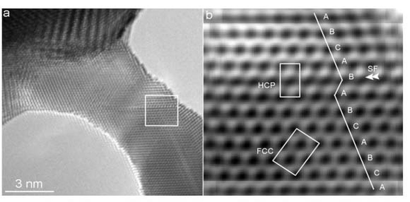
Those observations have led to foundational discoveries that are crucial for materials development. Scott X. Mao, MEMS professor, uses a specially designed sample holder to study how metals elongate and deform at the atomic level. Microelectronic mechanical systems rely on components made from microscopic structures of these metals, but metals behave differently at such a reduced length scale. Understanding the mechanical behavior of nanostructured metallic materials will enable the further development of strong and reliable components for advanced nanomechanical devices. Without such holder, it’s impossible to carry out an atomic scaled mechanical and electrical experiments under the most advanced high resolution electron microscope to achieve the understanding.
 Electron microscopy is used to observe
and test individual nanoparticles on flat surface in Jacobs' research.
Electron microscopy is used to observe
and test individual nanoparticles on flat surface in Jacobs' research.  Polymer with embedded copper
molecules.
Polymer with embedded copper
molecules.  Image from Roberts' paper
in ATVB, "Calcification in Human Intracranial Aneurysms Is Highly Prevalent and Displays Both Atherosclerotic and Nonatherosclerotic Types."
Image from Roberts' paper
in ATVB, "Calcification in Human Intracranial Aneurysms Is Highly Prevalent and Displays Both Atherosclerotic and Nonatherosclerotic Types." Anne Robertson, MEMS and BioE professor, and her team use the micro-CT in their NIH-supported work studying the causes for rupture of intracranial aneurysms (IAs). Robertson and her team used the specialized micro-CT equipment to analyze aneurysm tissue from patients and found that calcification is substantially more prevalent than previously thought. The micro-CT was able to identify microcalcifications as small as 3 micrometers. The team discovered differences in the types of calcification in ruptured versus unruptured aneurysms, made possible using the micro-CT system. The work was published in the journal Arteriosclerosis, Thrombosis, and Vascular Biology (ATVB (doi:10.1161/ATVBAHA.119.312922). This improved understanding could lead to new therapeutic targets and, ultimately, improved outcomes for patients with aneurysms.
Great innovations require the right tools. Thanks to Mr. Dudash’s gift, the MEMS Department has the tools to innovate, discover and create—tools that have produced an important base of knowledge that manufacturers will be building on for years to come.
“It is generous gifts from donors like Mr. Dudash that enable advanced research and, ultimately, discovery,” said Brian Gleeson, Tack Chaired Professor and MEMS Department Chairman. “Moreover, the funds provided by Mr. Dudash are being used strategically to create specialized capabilities that greatly help to procure further funding from agencies and, hence, further bolster research activities.”
Contact: Maggie Pavlick