
High field imaging with an ultra-high impact
7T MRI proven more effective than 3T, can reduce study cost and scale
Brain research is often limited by the realities of time, cost, and participant recruitment. Engineers at the University of Pittsburgh, however, have developed a way to ease these constraints: a uniquely powerful MRI system that delivers clear images and statistical significance while scanning just a fraction of the participants typically required.
“Neuroimaging studies are often limited by recruitment cost and feasibility,” said Tamer Ibrahim, professor of bioengineering at Pitt’s Swanson School of Engineering. “Reducing the required sample size shortens study timelines, lowers expenses, and broadens the types of questions that can be investigated.”
A publication in Human Brain Mapping from Ibrahim’s team, “Brain morphometrics correlations with age among 350 participants imaged with both 3T and 7T MRI: 7T improves statistical power and reduces required sample size” (doi.org/10.1002/hbm.70195), provides compelling evidence that ultra-high-field 7-Tesla (7T) MRI can substantially improve the detection of age-related structural changes in the human brain compared to scans with lower magnetic fields.
Evidence from this publication demonstrating 7T’s improved efficiency has strengthened investigations into menopause-related brain changes, mild cognitive impairment, and other aging-related conditions, allowing researchers like Rebecca Thurston, distinguished professor of psychiatry, clinical and translational science, epidemiology and psychology at the School of Medicine justify the use of 7T MRI in their grant submissions and research.
“7T MRI has been integral to answering the questions that I address in my work, which is focused on the brain changes of the menopause transition.” Thurston said. “For example, it is ideally suited to delineating the subtle brain changes of the early perimenopause that we are studying in our current $7.5M NIH-funded MenoBrain study.”
Building a better scanner
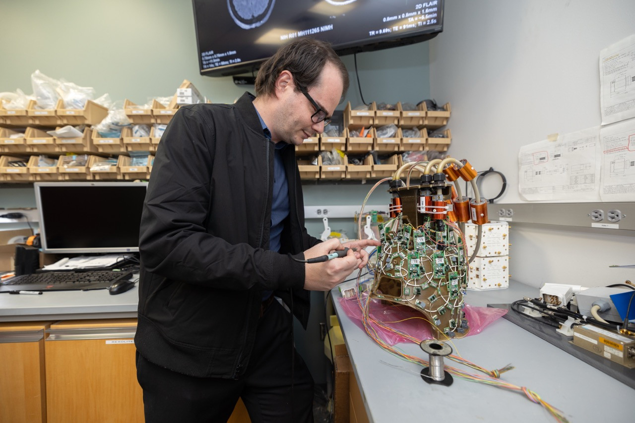
Magnetic resonance imaging (MRI) uses strong magnetic fields to create detailed images of the body’s tissues and organs. The magnetic flux density—measured in the unit Tesla (T) —helps determine the level of anatomical detail visible in the scan. While most MRI scanners operate at 1.5T or 3T, Ibrahim has developed specialized radiofrequency coils that allow Pitt’s 7T system to fully leverage this ultra-high magnetic field and deliver exceptional quality images.
“This publication shows that 7T provides more reliable and sensitive morphometric measurements when compared to 3T,” Ibrahim said. “For research questions involving subtle structural differences or longitudinal change in the brain, high performance 7T MRI is definitely the way to go.”
The large-scale, first of its kind study analyzed brain morphometry in 350 healthy adults between the ages of 29 and 68, each of whom completed imaging sessions at both 3T and 7T. The research team evaluated cortical and subcortical volumes, cerebral white matter, and mean cortical thickness. Across all measures, 7T demonstrated stronger correlations with age and revealed a greater number of brain regions, exhibiting more statistically significant age associations compared to 3T.
“Because 7T produces higher-contrast, lower-noise data, researchers need substantially fewer participants to detect meaningful effects with our 7T technology." Ibrahim said. “Studies that would require 350 participants at 3T could achieve statistical significance with approximately 100 participants at 7T.”
While 7T scanners exist at other institutions, their adoption for large-scale human studies has been limited since the magnetic strength can cause distortions and inhomogeneity in the images. Ibrahim and the 7 Tesla Bioengineering Research Program (7TBRP) have been troubleshooting these limitations for over the last 20 years by developing a custom radiofrequency coil system, Tic-Tac-Toe, which enables the 7T scanner to work smoothly and create the sharpest images possible.
“There are significant challenges when scanning at 7T. The interactions between the electromagnetic waves and tissue can lead to regions in the brain where there’s simply no MRI signal to detect.” Ibrahim said. “But our anti-claustrophobia Tac G2 coil system is, to my knowledge, the only one in the world that has successfully and comprehensively solved this problem. We don’t have those voids in our 7T images, and there are no barriers to running all types of MRI studies at 7T.”
Following the successful development of the first generation coil system, Tac G1, which was used on about 2,000 in-vivo human scans, the Tac G2 coil system implemented in 2022 has been used on more than 2,500 in-vivo human scans, exceeding the Tac G1’s usage in less than half of the time. This development has since enabled over 40 NIH-funded studies across aging, psychiatry, neurology, and cognitive neuroscience.
7T’s impact
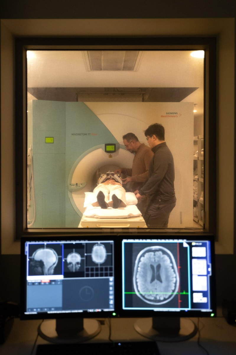
With these ongoing studies, many researchers and students at Pitt across medicine, psychology, engineering, and more utilize the 7T for their studies year-round, including bioengineering visiting research assistant professor Tales Santini and bioengineering graduate student Cong Chu, both working in the 7TBRP.
“As a young faculty member, I feel fortunate to have access to what is likely the largest paired 3T and 7T dataset, which has enabled us to directly characterize differences in brain structure measurements arising from these imaging techniques.” Santini said.
“And as a student, I'm excited to continue exploring this data to maximize the 7T's potential and to establish cross-platform harmonization methods for my future research.” Chu added.
For Anna Marsland, professor of psychology, nursing and clinical translational science at the Dietrich School of Arts and Sciences, the technology has supported her work through visualizing brain regions known to decline with age in the Adult Health and Behavior Cohort project, allowing her team to closely examine factors related to neurocognitive aging.
“The greater acuity of 7T imaging permits assessment of individual differences in the volume of subcortical brain regions, increasing our ability to identify brain regions that subserve cognitive functions and to identify individuals on accelerated aging trajectories who may be at increased risk for dementia.” Marsland said.
While each generation of coil systems can take about eight years to develop, working with researchers like Thurston, Marsland, and Santini and students like Chu to better understand the brain is exactly the kind of outcome that sustains Ibrahim and his team to keep improving the technology.
“When our coils are used in human studies, it’s incredibly rewarding, far more rewarding than just publishing this paper.” Ibrahim said. “We’re developing devices that clinicians and scientists use, and the result isn’t just pretty pictures; our engineering innovations are being used in real patient studies. We’re not making something that just could be used some time in the future, we are impacting human life now.”